STOMATOLOGY EDU JOURNAL 2022 Volume 9 Issue 1-2
CURRENT ISSUE
TABLE OF CONTENTS
GUEST EDITORIAL
Oral Health Systems in Europe – an overview
 Oral diseases are important components of noncommunicable diseases (NCDs). Notably, oral diseases afflict people of all ages. They often involve pain and discomfort, loss of oral functioning, impair quality of life, and they may lead to loss of work or school hours [1]. The predominant diseases and conditions of the mouth are dental caries, periodontal disease, loss of natural teeth, and oral cancer.
Oral diseases are often associated with the major chronic diseases because of common risk factors, primarily an unhealthy diet rich in sugars, use of tobacco, and excessive consumption of alcohol [2,3]. In addition, social determinants in oral health and health care are strong [1].
Oral diseases are a major public health problem to all countries in Europe. However, as for other NCDs, oral diseases vary extensively across countries and within countries. Social inequities in oral health status and use of oral health services are universal. Substantial variations in oral health care by education, family income and geographical areas are established among children, adolescents, adults, and older people throughout the European Region. Availability and access to oral health systems are important factors in people’s oral health status. The purpose of the present article is to outline the diversity of oral health systems in Europe; particularly, the variety of public and private involvement in health care is described. Additionally, the report provides important international statements on challenges to oral health systems development. The work is built on a regional survey carried out by the author among all European Chief Dental Officers 2018-2019. This survey was based on a World Health Organization structured questionnaire prepared for self-administration. Experts in oral health services research reviewed the final summary of answers.
(read more)
Poul Erik Petersen
DDS, Dr. Public Health Sci, BA
MSc (Sociology), Professor Emeritus
Editor-in-Chief Section
LETTER
Rudolf Slavicek’s Scientific Contributions
Jean-Daniel Orthlieb, Anne Giraudeau, Jean-Philippe Ré, Camille Raynaud, Florian Créhange, Estelle Casazza
Faculty of Odontology – Aix-Marseille University; APHM – La Timone Hospital, Marseille, France
 Rudolf SLAVICEK died in Vienna (Austria) on the first of January this year at the age of 93. He was serene, with a sense of accomplishment. He was an inspiring, tireless, strong-willed man, inventor and philanthropist. He devoted his life to the mastery of integrated occlusion in oral functions, acquiring a world reputation. His many scientific contributions will not only not fade away, but will grow: so, he will not disappear.
His history
Through his medical training (doctor of medicine in 1954, with incipient knowledge of cardiology), his training in dentistry (certified in 1957), his specialised training in restorative and prosthetic dentistry (1958), in orthodontics (1958-60), but also through his passion for anthropology and anatomy, Rudolf Slavicek gained an extremely broad cultural base, while in Vienna, Austria, between 1946, the year of his baccalaureate, and 1960. It is on this very solid base that he will build a professional career rich in innovations. Between 1960 and 1975 he initiated his quest for knowledge by reading and meeting the great international authors in the field of oral functions and dysfunctions. He worked, for example, with Lauritzen, Lundeen, Wirth, Gibbs and Ramfjord. Parallel to his private practice in Vienna, he developed a teaching career at a late stage, in which he demonstrated that the combination of broad culture, intelligence and a willingness to share can generate creative results that had a considerable influence on the field of occlusion, which concerned all aspects of dentistry.
He himself said “I took my time joining an academic career”. At the age of 50 (1978), he became an Associate Professor, defended his PhD in 1982, became a full-time University Professor in 1984, and was Dean of the Faculty of Dentistry in Vienna from 1992 to 1997. (read more)
ORIGINAL ARTICLES
- ORAL AND DENTAL DIAGNOSIS
DOI: https://doi.org/10.25241/stomaeduj.2022.9(1-2).art.1
 Biopsy is often an indispensable procedure in the diagnosis of myriad of benign and malignant oral conditions. The term “Biopsy” was introduced into medical terminology in 1879 by Ernest Besnier [1]. Biopsy is a procedure consisting of procurement of tissue from a living organism with the purpose of examining it under the microscope in order to establish a diagnosis [2]. The word biopsy originates from the Greek terms “bios” (life) and “opsis” (vision): vision of life [1,3].
Biopsy has been one of the oldest methods developed by the Arab physician Abulcasim (1103-1107AD), used for the accurate diagnosis of any abnormality in the oral environment as it is an accurate and pronominal aid used for establishing the histological characteristics of lesions which appear suspicious and so, it helps in their differentiation [4,5]. Biopsy of all kinds should be used frequently, not only for establishing initial/early diagnosis but also for providing more accurate clinical surveillance of the disease process. (read more)
Citation: Phulari RGS, Takvani MD, Vasavada D, Agrawal P. Attitude towards oral biopsy among general dental practitioners of Vadodara, a city in Western state of India. Stoma Edu J. 2022;9(1-2):15-20.
Authors:
Rashmi GS Phulari: ORCIDiD | ResearchGate | PubMed | Google Scholar
Mili D Takvani: ORCIDiD | ResearchGate | PubMed | Google Scholar
Dharmesh Vasavada: ORCIDiD | ResearchGate | PubMed | Google Scholar | Scopus
Prachi Agrawal: ORCIDiD | ResearchGate | PubMed
- DENTAL MATERIALS
Ex vivo digital comparison of four impression techniques using an industrial laser scanner
DOI: https://doi.org/10.25241/stomaeduj.2022.9(1-2).art.2
 One of the most critical steps in our dental processes is to make impressions with reasonable accuracy. The impression allows the dental technician to have the same condition on the model as in the patient’s mouth. When examining impressions, trueness and precision can be examined, and these two together constitute accuracy. ISO 5725 uses two terms, trueness and precision, to describe the accuracy of a measurement method. Trueness refers to the closeness of agreement between the arithmetic mean of a large number of test results and the true or accepted reference value. Meanwhile, precision refers to the closeness of agreement between test results [1]. In this study, only the trueness of the impressions was examined. A review article published in 2016 defined the still tolerable inaccuracy in the crown’s fit between 50 and 200 µm after the turn of the millennium [2]. The article by McLean and von Fraunhofer from 1972, which is still frequently cited, gives the 120-micron deviation as an inaccuracy threshold, so this level of accuracy must be aimed at for impressions [3-5]. (read more)
Citation: Jász B, Jász M, Körmendi S, Joós-Kovács G, Vecsei B, Hermann P, Borbély J. Ex vivo digital comparison of four impression techniques using an industrial laser scanner. Stoma Edu J. 2022;9(1-2):21-26
Authors:
Bálint Jász: ORCIDiD | ResearchGate |
Máté Jász: ORCIDiD | ResearchGate | PubMed | Scopus
Szandra Körmendi: ORCIDiD | ResearchGate | PubMed | Scopus
Gellért Joós-Kovács: ORCIDiD | ResearchGate | PubMed | Scopus
Bálint Vecsei: ORCIDiD| ResearchGate | PubMed | Scopus
- DENTAL MATERIALS
DOI: https://doi.org/10.25241/stomaeduj.2022.9(1-2).art.3
 There is abundant and strong scientific evidence that the appearance of a person’s face and teeth has a profound effect on perception and questioning by others [1–3]. It is also thought that the appearance of the face and teeth have a great impact on the development of the personality of the individual, getting a job, performing, believing in himself and being a victor. The social status of a personality and the attractiveness of a smile are related to each other [4].
While in the past, functional demands were taken into account in oral treatments, today the focus has shifted to aesthetic dentistry with the decrease in caries prevalence [5,6]. Establishing an appropriate balance between illusion and reality is the basis of aesthetic dentistry [7]. The ultimate purpose of aesthetic dentistry is to create beautiful smiles that are compatible with the teeth, gums, lips and face of the patient that complement each other in natural proportions [8]. One of the most important issues in aesthetic dentistry is color selection. Therefore, every dentist should know the color matching procedures for aesthetics [9].
For nearly a century, dentists have used tooth color shade guides for accurate color matching. This traditional way of picking colors is oversimplified and too subjective to constitute a standart [10]. While visual color selection with tooth color shade guides is the most common color matching system, it is considered inconsistent and subjective as it is affected by lighting, age, gender, eye fatigue [11]. In addition to the subject of color selection, which is a very challenging process in dentistry, dentists and technicians need to communicate about tooth colors during prosthesis production procedure. However, verbal communication of color differences is limited. A good color match is directly related to the quality of the prosthesis. The more precisely the tooth colors can be defined, the more accurate porcelain colors can be obtained [12–15]. (read more)
Citation: Ortaç D, Sonugelen M, Çömlekoğlu ME. Comparative evaluation of the relationship of teeth color and soft tissue color of the face in individuals with natural dentition. Stoma Edu J. 2022;9(1-2):27-37.
- DENTAL RADIOLOGY
Effects of cleft lip and palate on temporomandibular joint components: a CBCT study
DOI: https://doi.org/10.25241/stomaeduj.2022.9(1-2).art.4
 The temporomandibular joint (TMJ) is a complex joint located between the mandible and the temporal bone [1]. The loads applied to this joint affect both of the involved skeletal components, and can cause some alterations in their shape and thickness. In case of application of excessive forces, such alterations may exceed the normal range of variations (remodeling) and necessitate elimination of the etiology [2].
Cone beam computed tomography (CBCT) has gained popularity in recent years for imaging the craniofacial complex. CBCT delivers a significantly lower dose of radiation compared to conventional CT methods and has advantages over 2D images, including providing 1 : 1 orthogonal representations of structures. CBCT images can be used in the area of other 2D images, such as panoramic radiographic projection and lateral cephalogram, with the software capable of creating these images from the 3D data. Caution should be exercised to minimize radiation doses to patients. Studies have shown great variability in the amount of radiation exposure between different CBCT machines and the control of the field of view and intensity can help to minimize these levels. In addition, in cases with impacted teeth, CBCT images can provide a number of advantages over periapical and occlusal films for the localization of these teeth, since they provide images free of distortion and overlapping structures [3].
Approximately 60% to 70% of the populations worldwide show signs and symptoms of temporomandibular disorders; however, only one-fourth of them are aware of these signs and symptoms [4]. Temporomandibular disorders are
often characterized by pain at the TMJ, pain or tenderness of the muscles of mastication, mandibular movement limitation, mandibular deviation, and clicking of the TMJ. (read more)
Citation: Talaeipour AR, Kiaee B, Ghasemi S, Mirzaei A, Amiri F, Jamilian A, Darnahal A, Jamilian A. Effects of cleft lip and palate on temporomandibular joint components: a CBCT study Stoma Edu J. 2022;9(1-2):38-44.
Authors:
Ahmad Reza Talaeipour: ORCIDiD| ResearchGate | PubMed | Google Scholar | Scopus
Bita Kiaee: ORCIDiD | ResearchGate | PubMed | Google Scholar | Scopus
Shohreh Ghasemi: ORCIDiD | PubMed | Google Scholar | Scopus
Alireza Mirzaei: ORCIDiD | ResearchGate | PubMed | Scopus
Faezeh Amiri: ORCIDiD | ResearchGate | PubMed | Google Scholar | Scopus
Ayda Jamilian:
Alireza Darnahal: ORCIDiD | ResearchGate | PubMed | Google Scholar | Scopus
Abdolreza Jamilian: ORCIDiD | ResearchGate | PubMed | Google Scholar | Scopus
- PROSTHETIC DENTISTRY
DOI: https://doi.org/10.25241/stomaeduj.2022.9(1-2).art.5
 Edentulism represents a certain level of physical impairment, which is regarded as a chronic disability, causing many edentulous individuals to face obstacles in their everyday activities, such as eating or speaking [1]. Furthermore, it may significantly impact an individual’s psychological and social functioning and the overall quality of life [2,3,4]. Global trends have shown significant differences in the rates of edentulism worldwide [1,2,3,5]. Recent data from developed countries demonstrated a slight but encouraging decline in complete edentulism [6]. However, although edentulism is becoming less frequent in developed, industrialized countries, it remains prevalent in many parts of the world, and a complete denture is still one of the most frequent treatment options in cases of edentulism, especially considering older patients [1,6-9]. Prosthodontic treatment using a complete denture (CD) aims to achieve oral rehabilitation and reestablishment of the lost function, namely speech, occlusion, aesthetics, and masticatory function. It is considered one of the main challenges of prosthodontic treatment [9-15]. It is important to obtain satisfactory retention and stability of CDs, crucial factors for successful adaptation to them [15,16]. The masticatory function, antagonistic contacts, and preservation of masticatory muscle reflexes in CD patients were negatively correlated with the development of dementia in old patients [14,17]. The success of treatment with CDs is further influenced by many other factors that are also important to secure optimal retention and stabilization of a denture, such as characteristics of the saliva, status of the alveolar bone, condition of the mucosa and its resilience, relations between maxillary and mandibular residual alveolar ridges and neuromuscular factors [7-11,16,18]. Besides, a new denture may cause difficulties during speaking, and time is needed for patients to reach a satisfactory level of speech [16,18]. Although the quality of a new denture depends mainly on technological, biological, and physiological factors, it also depends on the interaction between the patient and the therapist [7]. (read more)
Citation: Poljak-Guberina R, Poklepović-Peričić T, Guberina M, Čelebić A. Duration and length of adaptation to new complete dentures: a survey based on patients’ self-reported outcomes. Stoma Edu J. 2022;9(1-2):45-53.
Authors:
Renata Poljak-Guberina: ORCIDiD| ResearchGate | PubMed | Google Scholar | Scopus
Tina Poklepović-Peričić: ORCIDiD| ResearchGate | PubMed | Google Scholar | Scopus
Marko Guberina: ORCIDiD| ResearchGate | PubMed | Google Scholar | Scopus
Asja Čelebić: ORCIDiD | ResearchGate | PubMed | Google Scholar | Scopus
REVIEW ARTICLE
- RESTORATİVE DENTİSTRY
Dentin degradomics in dentin erosion
DOI: https://doi.org/10.25241/stomaeduj.2022.9(1-2).art.6
 With the transformation of lifestyle dynamics and dietary habits, dental erosion has become an increased concern recently. Erosive tooth wear is an important oral health problem when considering the prolongation of human life and the survival of healthy dentition with the overall wellness approach. Regarding the ultraconservative dental concept, updated preventive strategies, and the recent technological improvements in the evaluating methods of enamel surface characteristics at both elemental and physical levels, dental researchers and clinicians have spent significant efforts to clarify the mechanisms of dental erosion. While only a few articles were available during the 1970s, today there are dozens of researches either in vivo or in vitro about dental erosion [1].
Dental erosion was previously defined as a sole substance loss by exogenous or endogenous acids without bacterial involvement. However, it was revealed in 2012 that dental erosion was not only a surface phenomenon but it showed a mineral dissolution beneath the surface [2-4]. It was proved that surface wear in the erosion process was heightened with the friction of acidic solution thus, dental erosion was not only a chemical dissolution but also a pathodynamic surface alteration [5]. (read more)
Citation: Ozan G, Berkman M, Sar Sancaklı H. Dentin degradomics in dentin erosion. Stoma Edu J. 2022;9(1-2):55-62.
PRODUCT NEWS
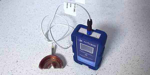
Prevention of peri-implantitis and bone recession using the Electronic Stem Generator
The scientific community recognizes there is no efficient treatment nowadays for the already installed peri-implantitis. As 34% of implanted pati-ents show peri-implantitis, the use of preventive treatments remains the only efficient approach.
Bone recession is the common disease in peri-implantitis, periodontal disease, grafts recession, periapical granuloma and orthodontic treatment. The cause of bone recession is the activation of osteoclasts, caused by microbes present in the oral cavity and occlusal forces. Scientific literature proves ratio receptor activator NF-kappa B ligand (RANKL) and its decoy receptor, osteoprotegerin (OPG) – RANKL/OPG as the bone recession indicator, which is higher as long as osteoclasts are activated.
In order to reduce the RANKL/OPG ratio, the certified 2A class medical device Electronic Stem Generator was created, based on long waves electromagnetic field to be applied in the patient’s home for a long time, and required for an efficient stimulation of deep bone progenitor cells in order to increase osteoblasts and OPG. (read more)
Florin – Eugen Constantinescu
DMD, PhD Student
Editorial Director, Product News
DOI: https://doi.org/10.25241/stomaeduj.2022.9(1-2).prodnews.1
BOOKS REVIEW
Short Implants
Editors: Boyd J. Tomasetti, Rolf Ewers
DOI: https://doi.org/10.25241/stomaeduj.2022.9(1-2).bookreview.1
Orthodontics and Periodontology
Combined treatments and clinical synergies
Authors: Roberto Kaitsas, Maria Giacinta Paolone
Publisher: Edra S.p.A., Milan, Italy
DOI: https://doi.org/10.25241/stomaeduj.2022.9(1-2).bookreview.2
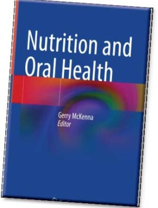
Nutrition and Oral Health
Editor: Gerry McKenna
Publisher: Springer Nature, Switzerland
DOI: https://doi.org/10.25241/stomaeduj.2022.9(1-2).bookreview.3
Infection Control in the Dental Office
A Global Perspective
Editors: Louis G. DePaola, Leslie E. Grant
Publisher: Springer Nature, Switzerland
DOI: https://doi.org/10.25241/stomaeduj.2022.9(1-2).bookreview.4


